• ACL Tears Can Be Repaired (Sometimes)

    By Dr. Derek Ochiai

    “A field hockey player is on a breakaway, with only one defender to beat before a clear shot to the goal. She makes a cut, and suddenly feels a pop in her knee, and she falls to the ground. The knee swells up, and the sports medicine doctor notes looseness in her knee. She gets an MRI, which shows an ACL tear, and the news is understandably devastating.”

    In the above scenario, typically the recommendation in 2017 would be rehabilitation to regain range of motion and strength in the knee. If the athlete wants to continue sports that involve change in direction and twisting, usually surgery is then recommended. Most of the time, ACL reconstruction (using a tendon graft) is the recommended surgical treatment. However, there are some ACL tears that can be repaired, strengthening and reinforcing the patient’s own ACL, with potentially a faster healing process.
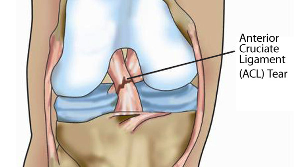
    What is the ACL?
    The ACL (anterior cruciate ligament) is one of the major stabilizing ligaments of the knee. It sits in the middle of the knee, and helps to control rotation.

    How does an ACL tear?
    Many times, ACL tears are non-impact, meaning that the athlete’s knee or body was not hit by another player or object. With quick change in direction, the stress on the ACL is overloaded, and a tear occurs. Other times, the athlete’s knee may be clipped by another, forcibly hyperextending the knee.

    What are the symptoms of an ACL tear?
    Initially, the athlete will usually fall or collapse because of the tear. Many times, they can leave the field under their own power (it is wrong to assume that an athlete who walks off under their own power does not have a significant knee injury). The knee may feel wobbly or unstable, or just “feel wrong”.

    How is it diagnosed?
    The athletic trainer or sports medicine physician will do specialized tests to see if the ACL is not working properly. The most common is a Lachman Test, which tests knee stability when the patient is relaxed and the knee is slightly flexed. The most specific test for ACL tear is the Pivot Shift Test, which tests the rotational stability of the knee (which is compromised by an ACL tear). Typically, X-rays are obtained to make sure there is no bony fracture of the knee. An MRI is usually obtained, which shows the torn ACL (Figure 1).
    Fig 1
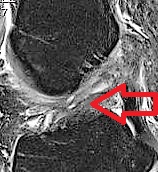
    Figure 1: MRI showing an irreparable mid-substance ACL tear. Notice how wavy the ACL is throughout the entire ligament.

    What are the treatment options for an ACL tear?
    Depending on the athlete and the mechanism of injury, some ACL tears can be treated conservatively, and the athlete feels OK. The ACL tear will not heal on its own, but the athlete can deal with the instability. For many athletes who want to go back to aggressive sports, surgery is usually recommended. ACL reconstruction is currently the gold standard for treatment of ACL tears. This involves a tendon graft used to replace the torn ACL. The graft sits in sockets in the femur and tibia, and the graft is held in place while it heals.
    Recently, ACL repair has emerged as an option for ACL tears that have torn off the bone of the femur. While not attached to the femur, the ACL itself has good tissue integrity, and if it heals, the ACL can function normally. If the tear is amenable to repair and if the injury is addressed relatively quickly, then ACL repair is an option.

    Fig2
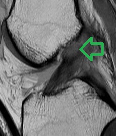
    Figure 2. This MRI shows a repairable ACL tear. Note how the black fibers are in continuity, and just avulsed off at the top.

    Why repair the ACL?
    ACL repair might be preferable to reconstruction for a number of reasons. First, repairing an ACL restores normal anatomy. While an ACL reconstruction makes the knee stable, it is using a tendon to recreate a ligament, which is not “normal”. The ACL has a blood supply and nerve supply, which can aid in proprioception. An ACL reconstruction graft does not have these. Finally, ACL repair may result in a quicker return to sport than ACL reconstruction.
    I thought ACL repairs didn’t work?
    Back in the 1960’s and 1970’s, surgeons would try to repair the ACL. However, the techniques used to “repair” are not anything close to what arthroscopic sports medicine surgeons have at their disposal now. Back then, repair was basically using low strength suture to re-approximate the ends of the ligament. There were no such things as suture anchors to repair ligament to bone. Modern ACL repair techniques take advantage of technology that has been used to successfully repair other tendons and ligaments, such as the rotator cuff. Remember, there were reputable doctors and scientists who contended it was impossible to run a four minute mile, and now high school runners are beating that mark!

    How do I know if my ACL tear is repairable?
    First, if you are feeling instability in your knee, you should be assessed by a sports medicine physician, to see if you have an unstable ACL. In this particular instance, it would be preferable to get an MRI relatively quickly, since ACL repair is better done in the acute setting (chronic ACL tears will result in tissue degeneration and scarring down of the stump of the ACL, making ACL repair either more difficult or not an option anymore). The MRI should be read by a sports medicine surgeon who could possibly repair the ACL, since just reading a report that says “torn ACL” does not indicate whether or not the ACL tear is repairable.

    Fig3
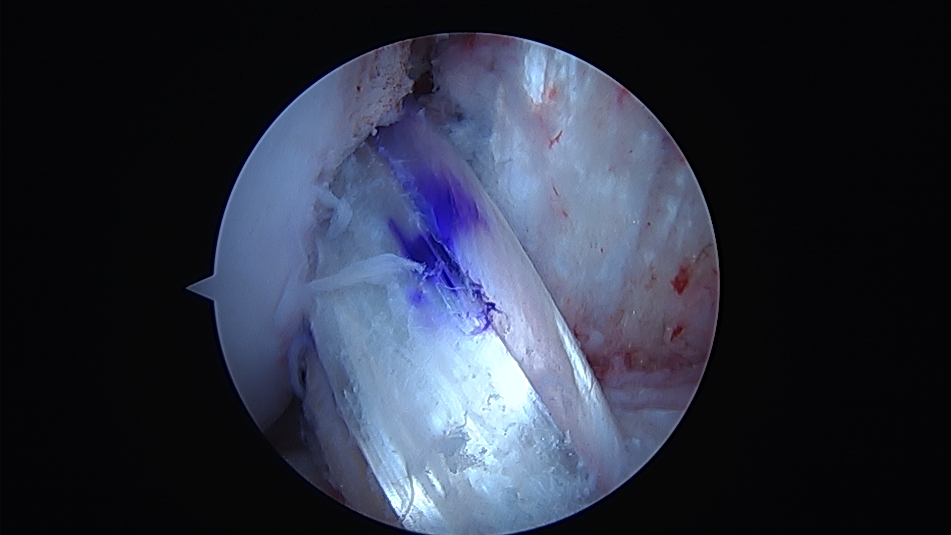
    Figure 3. Arthroscopic image of an ACL reconstruction using a hamstring tendon graft.

    Fig4
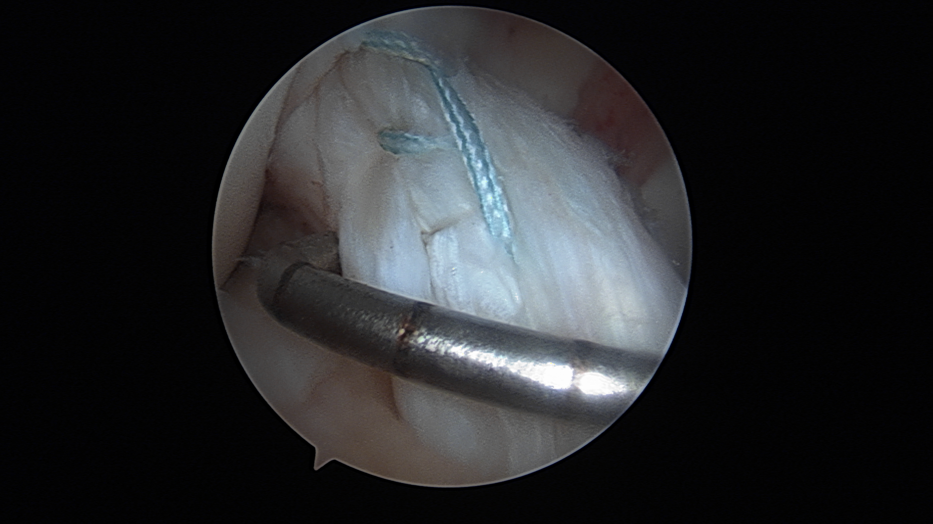
    Figure 4. Arthroscopic image of an ACL repair using suture anchors.

    In conclusion, many ACL tears in athletes require arthroscopic surgery. In 2017, the options for surgery could be either ACL reconstruction or ACL repair.

    Dr. Derek Ochiai is a Hip Arthroscopy and Sports Medicine surgeon at the Nirschl Orthopaedic Center. Follow him on Twitter: @DrDerekOchiai

  • What’s Up, Doc??

    51Kby4VZyQL._UY395_
    My physical therapists are a DPT. Does that mean they are doctors?

    Yes, but not medical doctors. They are Doctors of Physical Therapy. All DPTs must complete four years of an undergraduate degree, followed by an additional three years of graduate school. The DPT is a clinical doctorate. This means that, while in school, students apply their knowledge in rigorous, supervised internships in various clinical settings.
    The Doctor of Physical Therapy (DPT), is the standard degree for all physical therapists graduating after January, 2015. Since the transition to the status of a doctoring profession is quite recent, you may have a physical therapist that has a master’s or bachelor’s degree. No matter what the level of degree, all licensed physical therapists are trained to provide excellent and compassionate care to patients.

    Can DPT’s write prescriptions?

    No. Your physical therapist cannot write a prescription for any medications or order any diagnostic testing.

    What is the difference in schooling between a DPT and a PT?
    Prior to the DPT, the training for a physical therapy license required 2 years after graduating from college. The DPT is a three- year program and includes additional courses and longer supervised internships. The additional courses include advanced training in:
    • diagnostic and medical screening
    • diagnostic imaging
    • pharmacology
    • health care systems
    • business and economics
    • clinical research
    • health promotion and wellness
    • leadership and professional issues

    What does this mean to the patient?

    The DPT program prepares physical therapists to treat patients, and when needed to, refer them to other appropriate medical professionals such as orthopaedic surgeons. Currently 47 out of 50 states (including Virginia) allow DPTs to be direct access practitioners, meaning you can schedule an appointment with a DPT without first seeing a physician to obtain a written prescription for physical therapy. This greatly reduces the waiting time for people with injuries to get in and seek medical attention allowing for faster treatment and healing.
    Clinics, like Virginia Sportsmedicine Institute, that are committed to hiring DPTs send a message of dedication to best practices to their patients.

    This week’s blog we welcome guest blogger Dr. Skye Donovan, Associate Professor and Department Chair in the Physical Therapy program at Marymount University.
    faculty_donovan_skye

  • Foot or Knee Pain Orthotic Shoe Inserts May Help

    Custom molded orthotics are corrective shoe inserts designed specifically for each individual patient. They support and cushion the foot and improve biomechanics by putting the foot in correct alignment at every stage of walking, running and pivoting.

    ortho

    Orthotic inserts are often very effective in relieving many common complaints including:

    • Achilles Tendonitis
    • Aligning and Supporting the Foot or Ankle
    • Ankle Sprains
    • Haliux Rigid/Toe Pain
    • Heel Spurs
    • Hyper Pronation
    • IT Band Pain
    • Knee Pain
    • Leg Length Discrepancy
    • Low Back Pain
    • Metatarsalgia
    • Neuroma
    • Pes Planes/Flat Feet
    • Plantar Fasciitis
    • Rigid/Supinated Foot
    • Sesamoiditis
    • Shin Pain
    • Tendonitis

    For example, a wedge inserted into the inner (medial) side of the sole of a shoe can be used to help support a Pes Planus/flat foot, thus reducing the risk of tendinitis. A heel flare inserts can be used to increase support and help prevent ankle sprains. Heel cushions can help absorb impact and relieve stress on the heel and ankle when you walk or run. The type of orthotic recommended will depend on several factors such as; your symptoms, the underlying cause for those symptoms, your comprehensive evaluation, the type of activity/sports you perform, and the shape of your feet.

    screen

    Although custom orthotics are considerably more expensive than off-the-shelf devices, they last much longer and provide better more individualized corrective support. In some cases, an over-the-counter device can be just as effective when combined with a stretching and exercise program. Here at Virginia Sportsmedicine Institute a comprehensive evaluation, including gait analysis, muscle and flexibility testing, and a computerized foot analysis is used to develop your orthotics. The type of orthotic recommended for you will depend on several factors including: the underlying cause of the symptoms you are experiencing; the type of activity/sports you perform; the shape of your feet. The following are the main types of custom shoe inserts:

    Rigid Orthotics– Made of firm materials like plastics, fiberglass, and carbon fiber; these primarily address motion in the two major foot joints below the ankle. If a foot imbalance is the cause of pain in your legs, thighs or lower back, rigid orthotics generally are recommenced. These are usually designed for use in walking shoes or dress shoes.

    Semi-Rigid Orthotics– These act as a balance aid when walking or performing athletic activities. They are made from foam with a thin, plastic shell laminated to it. These devices provide accommodative support while allowing the foot to be the mobile adapter.

    Soft Orthotics– Designed to act as shock absorbers and also to increase balance and reduce pressure in sore spots, these inserts are made of compressible foam. They have proven effective for arthritic feet and are widely used to care for diabetic foot conditions. They tend to be bulkier than other inserts and may require extra room in shoes or prescription footwear.

    If you think you may benefit from orthotics, check with your doctor to get a prescription and call our Virginia Sportsmedicine Institute at 703-525-5542 to set up an evaluation. It is important to bring in the shoes you regularly wear so we can check the wear pattern and stability of your shoes. In addition bring in any orthotics or shoe inserts you may have used or currently use. Each insurance policy is different so it is important to check with your specific insurance carrier on coverage. We can provide you with the procedure codes to help the process.

  • Pain Relief Fast with Trigger Point Dry Needling

    What is Dry Needling?
    Dry needling is a treatment technique used by physical therapists to eliminate trigger points within muscles. Trigger points are small knots of tightly contracted muscle that are often sore to the touch. These “muscle knots” can cause pain, limit motion and affect performance. If left untreated, they can worsen over time. In dry needling, a sterile thin filament needle is inserted into the trigger point, causing it to twitch or “release”. This release reduces pain and muscle tension, and increases mobility.
    Dry needling does not take the place of other hands-on therapy or exercises, but it is a valuable treatment option. Dry needling, combined with other physical therapy treatments, can help the following conditions:

    Acute and chronic tendonitis
    Athletic and sports-related overuse injuries
    Carpal tunnel syndrome
    Chronic pain conditions
    Ehlers Danlos Syndrome
    Frozen shoulder
    Fibromyalgia
    Groin strains
    Hamstring strains
    Headaches and whiplash
    Hip pain
    Knee pain
    Lower back pain
    Muscle spasms
    Plantar fasciitis
    Post-surgical pain
    Post-traumatic injuries, motor vehicle accidents, and work related injuries
    Repetitive strain injuries
    Sciatic pain
    Tennis elbow
    Many other musculoskeletal conditions . . .

    How Long Does it Take for Dry Needling to Work?
    In some cases, decreased pain and improved mobility is immediate. Typically, it may take a few treatment sessions for a lasting positive effect.

    What are the Advantages of Dry Needling?
    Access – The advantage over other techniques is that we can treat parts of the muscle and deeper layers of muscles which our hands and fingers cannot reach, and it works faster than massage at relaxing the muscles.
    No Drugs – There are no drugs used in dry needling, so we can treat many trigger points during each treatment.
    Immediate Relief – Deactivation of the trigger points can bring immediate relief of symptoms, and then we can immediately stretch and train the muscles to work in their new pain free range of motion. Thus, results are achieved with dry needling which cannot be obtained with any other treatment.

    Will Dry Needling Help Me?
    Does a massage give you great relief, but the relief doesn’t last as long as you would like? People who have good results with massage, but are disappointed when the discomfort returns, will find dry needling a great way to get more long-lasting relief. We are able to treat almost any muscle in the body, and treat the muscle at depths impossible with other types of bodywork. Dry needling is a great way to get more out of your physical therapy by allowing us to eliminate the deep knots and restrictions that have, up until now, been unreachable.

    How Many Needles Will I Need?
    We will start very slowly during the first session to give you a feel for the technique. The first session will focus on a few muscles that are key to your problem. These key areas can give you excellent relief with less soreness. Subsequent treatments will target more specific areas to fine-tune the effect. Sessions are usually spaced 5-7 days apart and you should expect to feel a marked difference after only 1or 2 sessions.

    How Will I Feel After Dry Needling?
    You will know positive change has occurred right after the session, because you will be sore in the way that you would feel after increased activity. The muscle will feel fatigued, and the soreness can last from a few hours to 1 or 2 days, but should not interfere with your everyday activities. We encourage you to be active during this time to keep the soreness to a minimum. After a day or so, you’ll experience a new and lasting feeling of less pain and tightness. The injury and pain you thought was there to stay will actually start to diminish.

    Call our office today to make an appointment or For more information. Call Virginia Sportsmedicine Institute at 703-525-5542 ext. 200, or go to our web site www.vasportsmedicine.com.

    dry-needling

  • The Power of Determination- Life As A One-Handed Gymnast

    As a 17 year-old high school senior, Leah qualified for her first Level 9 all-round Virginia State gymnastic competition. Just two months before the big event, she under rotated on a vault and fell directly on her left hand. She knew immediately that something was wrong from the significant bruising and pain.  At the office of Dr. Cassie Root, hand surgeon at the Nirschl Orthopaedic Center, her x-rays showed a displaced spiral fracture of the 5th metacarpal.

    Dr. Root discussed all the options with Leah and her family and Leah decided to proceed with surgery. Two days later, Dr. Root performed a closed reduction and placed two wires to hold the fracture in the proper alignment. A few days later, Leah was placed in a custom splint fabricated by Meghan Little, a certified hand therapist at Virginia Sportsmedicine Institute.  After a serious discussion with Dr. Root, it was agreed upon that Leah could continue her gymnastics but with one major condition. She could only use one hand! Three weeks after surgery, Dr. Root removed the wires in the office.  Leah had regained almost all of her finger range of motion due to her hard work in physical therapy with Meghan.

    gymnastics

    On her final visit to the doctor only 4 days before the gymnastic competition, the x-rays looked great. Leah was allowed to slowly decrease the use of her splint but she could not yet tumble on her injured hand.

    Of course this meant Leah could not compete in the all-round events, but she loved the floor routine.  This would be her last meet as a senior.  So, with only 3 weeks until States, she completely changed 3 vital skills in her floor routine so she could compete. If you have ever been a competitive athlete, you can appreciate how challenging it is to completely change your routine only 3 weeks prior to a competition. To top that off, she could only tumble using one hand. This was a once-in-a-lifetime opportunity for Leah, and she had worked for many years to accomplish this goal.

    Leah qualified for Regionals and competed her one arm routine again. Although she did not place at Regionals, Leah was just so happy to compete in the state competition. Her grit and determination should be a lesson to us all. Sometimes when life throws you a curve ball, pick it up and throw it right back – even if it is with only one hand!

    Nirschl Orthopaedic Center is a leader in sports medicine and general orthopedics services. If you have an orthopaedic injury, schedule an appointment with one of our doctors today by calling our Arlington, Virginia location at 703-522-2200. Visit our website at www.nirschl.com to learn more about our services. Follow us on Twitter @DrCassieRoot and @NirschlOrtho.


  • The top 10 tips to enjoy Thanksgiving (and have your body thank you for it)

    thanksgiving

    Thanksgiving is around the corner, and everyone’s looking forward to getting together with family and indulging (why not?). Fortunately, we have put together 10 ways for you to ‘have your turkey and eat it too.’
    Here they are:

    1. Drink plenty of water throughout the week – especially before heavy meals.
    2. Walk off that big dinner (you can even make it a family outing and exercise while enjoying one another’s company.
    3. Create a caloric deficit by upping your exercise routine the week of the big meal.
    4. Make simple substitutions, such as lighter milk and cream, to enjoy your favorite treats without the heavy health impact.
    5. Make small cutbacks on sugars and salts involved in the cooking process as most recipes call for more than is needed.
    6. Continue to have evenly-paced meals throughout the day. Having a normal breakfast and lunch will help you to refrain from overdoing it at dinner and dessert.
    7. Use smaller plates. Many times people will overeat purely because they pile larger portions onto wide plates.  Give yourself a mental edge by using a smaller plate and having smaller portions.
    8. Eat slowly to allow your body time to begin digestion. You may find yourself feeling full far earlier than you would have thought.
    9. Beware of alcohol intake. It’s easy to drink more than your normal amount during holidays, but too much alcohol can cause a variety of problems, including dehydration.
    10. Relax. Stress is a huge health risk and contributes to a vast variety of ailments.  Allow yourself to enjoy the holiday.

    From all of us at Nirschl Orthopaedic Center and Virginia Sportsmedicine Institute Physical Therapy we wish you a very Happy Thanksgiving!

  • Traveling For Surgery?

    SurgicalTravel-150x150

    By Derek Ochiai, MD

    Our society is becoming more global and travel friendly.  Many times, patients with a specific medical issue may decide to travel out of town to have definitive medical treatment, including surgery.  While there may be very good reasons to want or need to do this, the patient may have additional issues and needs that must be considered prior to making this commitment.  As a surgeon who often sees patients who travel from out of town for a specialized procedure that I do, I am well aware of these issues.  I have currently operated on patients from California, Idaho, Iowa, Michigan, North Carolina, Oklahoma, Pennsylvania, Puerto Rico,  and Texas. This article will talk about what to ask and look out for when traveling for surgery.

    Patients have reasons that they wish to travel to have surgery.  The doctor or center that they are choosing may have a specific expertise for a medical condition.  The patient may also travel to be closer to their family to make for an easier recovery.

    There are four specific issues that a patient should consider prior to having surgery out of town.  First, how long can the patient stay away from home?  While a surgery may normally take a set amount of time for initial recuperation, the surgery may not be without complications.  If the surgeon wants you to stay an extra week after the procedure, is that personally and financially feasible?  Second, what happens if there is a late complication after the patient returns home?  The surgeon may request that you return for follow-up care and this can be an additional expense and time away from home and work.  If you cannot get back to that surgeon, are their other physicians local to you who would be willing and able to treat you?  Third, how communicative is the surgeon?  Many orthopaedic procedures rely on physical therapy and post-surgery protocols after surgery.  If the patient or other health care providers have questions regarding this, how does this get communicated to the surgeon?  Does he/she answer email?  Is the surgeon comfortable with treating a patient “over the phone”?  Finally, how comfortable is the surgeon treating a patient whom he/she will not be following long term?  Do they have a system in place to handle issues of communication and follow-up?

    The most important step a patient can take is to research doctors and facilities ahead of time.  Many times, doctors who are accustomed to seeing patients from out of town have a system in place to accommodate this. The patient can gather their medical records, radiographic images and MRI’s, which can be mailed ahead of time for the surgeon’s review.  If a patient is thinking about having their surgery performed out of town, a call to the surgeon, asking if this system is in place, can save some unnecessary expense.  It is far better for patients to know ahead of time if they are an appropriate candidate for a procedure, rather than travel to the facility only to find out otherwise.  A patient should also ask to speak to the billing department, as the center and surgeon either may not participate with their insurance or the specialized procedure may not be covered by their insurance.  By talking to the staff, the patient can definitely get a sense of how experienced the office is in dealing with patients from out of town, which can help with the patient’s comfort level.

    All patients should set up a pre-operative appointment well before an intended surgery.  This will relieve pre-surgery anxiety for the patient who now knows their doctor prior to having the surgery.  Sometimes, no matter how great a doctor looks on the internet or how convenient it would be to have the surgery there, a patient may meet the surgeon and decide to look elsewhere.  If the pressure of already scheduling the surgery is looming, this may unduly influence an important decision.  Keep in mind that many centers may also have a surgical cancellation fee.

    Medical tourism has its benefits and drawbacks.  If a patient actively researches and prepares before surgery, the benefits can be maximized, and the drawbacks diminished.

    Dr. Derek Ochiai is a board certified orthopaedic surgeon, specializing in Hip Arthroscopy and Sports Medicine, at Nirschl Orthopaedic Center for Sports Medicine and Joint Reconstruction.  Follow him on Twitter @DrDerekOchiai. For more information go to the web site at www.nirschl.com.

    Nirschl Orthopaedic Center is a leader in sports medicine and general orthopedic services. In addition, Virginia Sportsmedicine Institute Physical Therapy has been rated on of the top sports medicine clinics in the Washington DC metropolitan area. If you have an orthopedic injury, schedule an appointment with one of our doctors today by calling our Arlington, Virginia office at 703-522-2200 or visit our websites to learn more about our services.


  • Hip Labral Tears and Femoroacetabular Impingement 

    Derek Ochiai, MD

    Nowadays, it is rather commonplace to read about an athlete undergoing hip arthroscopy for a labral tear of the hip. This article will review what a labrum is, why it’s important, how it tears, and what kind of treatment is available.

    What is a labrum?

    The labrum is a rim of cartilage that surrounds the hip joint. It attaches to the socket of the acetabulum. If you have friends who had a knee arthroscopy for “torn cartilage”, they had a meniscal tear of the knee. The meniscus of the knee and the hip labrum are made up of the exact same type of cartilage.


    Figure 1: Arthroscopic picture of a normal posterior labrum. The labrum attaches smoothly to the acetabular articular cartilage. The femoral head is on the bottom right of the picture.

    Why is the labrum important?

    The labrum increases the relative depth of the socket, which can confer additional stability to the hip. This can be especially important in ballet dancers, figure skaters, gymnasts, and other athletes who put their hips through extreme ranges of motion. In developmental dysplasia of the hip (DDH), the socket is much shallower than normal, and the labrum deepens the socket to keep the femoral head of the hip in that shallow socket. Also, there are medical conditions where a person has increased joint laxity (such as Ehlers-Danlos), where the hip is more unstable and relies on the labrum for stability.

    The labrum acts a seal around the femoral head, to maintain fluid pressure of the hip joint.

    What are the symptoms of a labral tear?

    The severity of symptoms can vary. The “hip pain” 90% of the time is perceived as deep in the groin. Sometimes, the pain can radiate to the side or the back of the hip as well. Many times, patients with a labral tear have pain and/or a feeling of catching in their hip, especially when going from sitting to standing. They may notice that they have to compensate to get in and out of cars. They may have pain with squatting and exercise, especially with sports that involve cutting and changing direction. Many times, patients also complain of pain with sex.

    I didn’t injure my hip. Why do I have a labral tear?

    The vast majority of labral tears are from FemoroAcetabular Impingement (FAI). FAI is a condition that develops in a person’s early teen years, where the hip is “out of round”. Since the hip joint and labrum are meant to function with round on round mechanics, this out of round conflict puts increased stress on the labrum. Over time, this increased stress can cause the labrum to tear. Symptoms of FAI greatly overlap with labral tears. In addition, symptomatic FAI can cause pain with prolonged sitting.

    How is a labral tear diagnosed?

    A medical professional can suspect a labral tear based on a patient’s symptoms and history. Clinically, the doctor can do provocative tests, such as the anterior and posterior impingement test, the McCarthy test, and the FABER exam, to further investigate. Many times, X-rays are very useful. FAI is a radiographic diagnosis, and usually can be easily seen on plain X-rays. With severe FAI, a labral tear can be inferred.


    Figure 2: On left side of screen, normal acetabulum. The anterior wall (red line) and posterior wall (blue line) do not cross. On right, there is pincer type FAI, where the red and blue lines cross.


    Figure 3: Typical cam type FAI X-ray finding. The yellow outline shows what the contour of a normal hip would look like.

    MRI (magnetic resonance imaging) directly shows the cartilage of the hip. While a labral tear can be diagnosed with a plain MRI, an MRI arthrogram is more sensitive to labral tears. An MRI arthrogram does involve an injection directly into the joint, but then the dye can easily be seen leaking into a labral tear, making the diagnosis clearer.


    Figure 4: MRI arthrogram of a left hip labral tear. Arrow points to the dye leaking between the labrum and the articular cartilage.

    I’ve been diagnosed with a labral tear. What do I do?

    The mainstay of initial treatment for hip labral tears is physical therapy and activity modification. Many patients ask me how this is going to “cure” their labral tear. Physical therapy can help improve core/gluteal strength, which can shift the femoral head back in the socket a bit. This can decrease stress on the labral tear, which is normally near the front of the socket. While this doesn’t “cure” a labral tear, it can make some patients feel significantly better. Sometimes, your doctor may suggest an intra-articular cortisone injection to the affected hip. While this also does not “cure” a labral tear, it can sometimes act as a physical therapy aid, allowing patients to “get over the hump” with initial therapy and start building core/gluteal strength.

    All that didn’t work. My surgeon says I need a hip scope.

    Labral tears and hip FAI can now be addressed by hip arthroscopy, using cameras and small instruments inside the hip. “Scoping the hip” means looking inside the joint; there are multiple possible procedures that could potentially be performed during hip arthroscopy. In the past, the most common procedure was labral debridement, or trimming out the torn labrum. While this has the advantage of not relying on the body to heal a labral repair, several studies have shown that labral repair has better long term outcomes than debridement. At least in my practice, labral repair is much more common. The labrum is repaired by drilling anchors into the bone of the socket, and using its sutures (thread) to wrap around and through the labrum to tie the labrum back into place. When doing a labral repair, any FAI should be addressed at the same time. Otherwise, there is a good chance of the repair failing (because the forces that tore the labrum would be the same forces causing it not to heal). Sometimes, a surgeon may tell a patient that the best procedure is an open surgical dislocation (through a large incision). There are some special cases where this approach may be preferable, but it is not common.


    Figure 5: Large anterior labral tear. Note the separation between the labrum and the acetabulum. Compare this to Figure 1.


    Figure 6: Picture of a labral repair. In the picture, there are three sutures that are anchored to the bone, sewing the labrum back to the acetabulum.

    A newer procedure to address labral tears is labral reconstruction. This uses a tendon graft to take the place of the torn labrum. Typically, this is only used for labrums that are so torn and degenerative, that repairing the labrum will not work to restore normal labral function.


    Figure 7: Labral reconstruction: In this picture, the labrum was not repairable. Instead, a tendon was used as a graft, to reconstruct the labrum.

    Hip arthroscopy is a passion of mine. I hope this has been helpful to you. If you have been diagnosed with a labral tear, please ask your surgeon specific questions. Over the years, I have seen many second opinions from patients who had less than successful outcomes from hip arthroscopy that thought they had a labral repair, but the surgeon actually did a labral debridement. To me, “fixing a tear” means sewing it back and anchoring the labrum; however, “fixing a tear” to some could also mean “fixing the problem caused by the tear”, which could mean debridement.

    Dr. Derek Ochiai is a board certified orthopaedic surgeon, specializing in Hip Arthroscopy and Sports Medicine, at Nirschl Orthopaedic Center for Sports Medicine and Joint Reconstruction. Follow him on Twitter @DrDerekOchiai. For more information go to the web site at www.nirschl.com.

  • What Is Good For Your Heart Can Be Murder On Your Elbow

    Dr. Robert Nirschl on televisions Good Morning America, with Charlie Gibson, discuss preventing tennis injuries. This interview was aired in 1996 but is still very relevant today.